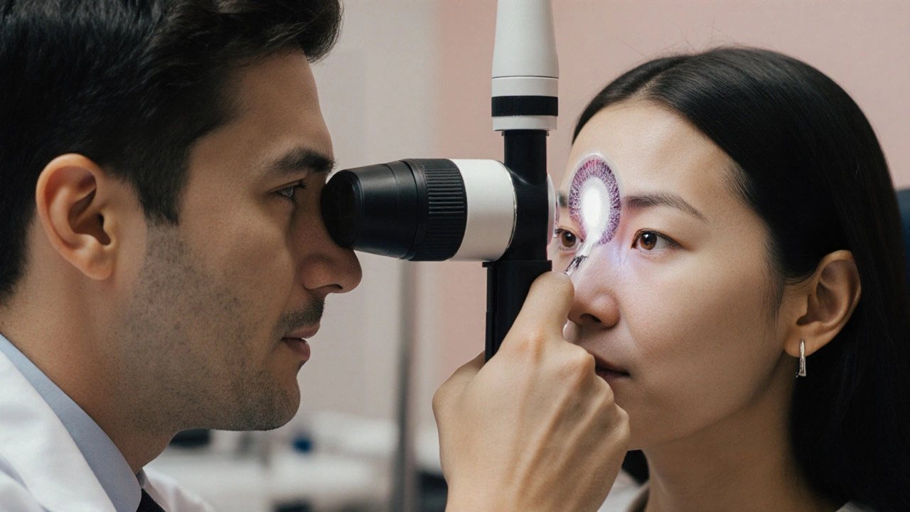
Eye Infection Symptom Checker
This tool helps identify possible eye infections based on pupil behavior and symptoms.
Ever wondered why your eye sometimes goes dark when it’s sore or infected? That tiny change is called myosis - a medically‑sound term for pupil constriction - and it can be a hidden clue in many eye infections and inflammatory conditions.
TL;DR
- Myosis = pupil gets smaller; it’s controlled by the parasympathetic nervous system.
- Inflammation releases chemicals that trigger myosis as a protective reflex.
- Uveitis, conjunctivitis, and keratitis often show a constricted pupil, while severe corneal ulcers may keep it dilated.
- Doctors use myosis to gauge infection severity and choose treatments.
- If you notice a sudden, painful constriction, get checked - it could signal a serious infection.
What Exactly Is Myosis?
Myosis is the medical term for a reduction in the pupil’s diameter. It’s the opposite of mydriasis (pupil dilation). The key attributes are:
- Cause: activation of the parasympathetic fibers of the autonomic nervous system.
- Typical size: 2-4mm in bright light, expanding to 4-8mm in darkness.
- Function: limits light entry, protects the retina, and enhances near‑vision focus.
When an infection or inflammation hits the eye, this reflex can kick in automatically, “closing the blinds” to shield delicate structures.
How the Iris and Autonomic Nervous System Pull the Strings
The colored part of the eye - the iris - houses two muscle groups:
- Sphincter pupillae: contracts under parasympathetic influence, producing myosis.
- Dilator pupillae: relaxes under sympathetic input, causing dilation.
Inflammatory mediators like prostaglandins and cytokines stimulate the parasympathetic pathway, tightening the sphincter. In simple terms, the eye’s built‑in “shutter” snaps shut when it senses trouble.
Why Infections Trigger Myosis
When bacteria, viruses, or fungi invade the ocular surface, the immune system releases a cascade of chemicals:
- Prostaglandin E2 (PGE2): directly enhances sphincter muscle tone.
- Acetylcholine: the main messenger of the parasympathetic nerve, causing rapid constriction.
- Histamine: increases vascular permeability, adding to swelling that mechanically pushes on the iris.
These agents act together to shrink the pupil within seconds to minutes, a protective reflex that reduces glare and limits pathogen spread.

Common Eye Infections & Their Typical Pupil Responses
| Infection | Typical Pupil Reaction | Underlying Reason |
|---|---|---|
| Conjunctivitis (bacterial or viral) | Mild to moderate myosis | Inflammatory cytokines stimulate parasympathetic fibers. |
| Uveitis (anterior) | Prominent myosis | Severe intra‑ocular inflammation → high prostaglandin levels. |
| Keratitis (corneal ulcer) | Variable - often normal or mild dilation | Pain dominates, activating sympathetic response. |
| Endophthalmitis (post‑surgical) | Irregular - may show sluggish myosis | Combined inflammation and intra‑ocular pressure changes. |
| Herpes Simplex Keratitis | \nInitial myosis, later possible dilation during ulceration | Early immune response then nerve damage. |
Clinical Significance: Using Myosis to Diagnose
Eye doctors (ophthalmologists and optometrists) treat pupil size like a traffic light. A sudden, painful constriction often signals:
- Acute anterior uveitis - requires corticosteroid eye drops within hours.
- Severe bacterial conjunctivitis - may need antibiotic ointments.
- Potential intra‑ocular pressure spikes - a red flag for glaucoma.
Because myosis appears before redness or discharge in many cases, catching it early can speed up treatment and protect vision.
Treatment Implications: When Myosis Helps, When It Hinders
Medications that influence pupil size are double‑edged swords.
- Pilocarpine: a parasympathetic agonist that deliberately induces myosis; used to treat acute angle‑closure glaucoma but can worsen infections by limiting drug penetration.
- Atropine or cycloplegics: block parasympathetic signals, causing dilation. In uveitis, doctors may use them to “rest” the iris and reduce painful spasms.
- Topical NSAIDs: lower prostaglandin production, indirectly easing myosis and pain.
Understanding the pupil’s role lets clinicians pick the right eye drops, avoiding a scenario where a drug intended to relieve pressure actually fuels an infection.
Practical Tips for Anyone Experiencing Myosis
- Note the timing - does the constriction happen suddenly after eye pain or redness?
- Check for accompanying symptoms: excessive tearing, light sensitivity, or blurred vision.
- Avoid eye rubbing - it can spread microbes and worsen inflammation.
- If you wear contact lenses, remove them immediately and switch to glasses until a professional evaluates you.
- Seek prompt ophthalmic care if myosis is painful, asymmetrical, or accompanied by visual loss.
Frequently Asked Questions
What causes myosis without an infection?
Bright light, certain medications (like opioids), or a normal parasympathetic response during near‑vision tasks can all shrink the pupil without any disease.
Can myosis be a sign of a serious eye condition?
Yes. Sudden, painful constriction often points to anterior uveitis or acute angle‑closure glaucoma - both need urgent treatment to prevent permanent vision loss.
Is myosis contagious?
No. Myosis is a reflex, not an infectious agent. However, the underlying infection that triggers it can be contagious (e.g., viral conjunctivitis).
Should I use over‑the‑counter eye drops if my pupil is constricted?
Avoid self‑medicating. Some OTC drops contain vasoconstrictors that may mask symptoms or worsen inflammation. A professional exam is the safest route.
How long does infection‑induced myosis last?
It typically resolves as the inflammation subsides - anywhere from a few hours to several days, depending on treatment effectiveness.


Christopher Ellis
September 29, 2025 AT 18:31It seems the eye is a tiny lighthouse and myosis is its way of screaming that the seas are stormy yet we ignore the warning because we prefer the bright glare of ignorance
kathy v
September 29, 2025 AT 18:33As an American who cares deeply about the health of our nation, I have to point out that the information presented here is a perfect example of why we need more rigorous standards in medical education. The description of myosis is thorough, but it fails to emphasize the urgency that our citizens must feel when faced with potential eye infections. We cannot sit back and let people ignore a painful pupil constriction while scrolling through memes. The United States has some of the best ophthalmologists in the world, and we should be proud to share that expertise, not hide behind vague explanations. Moreover, the mention of prostaglandins and cytokines could be expanded to include how our healthcare system provides access to the latest anti‑inflammatory treatments. It is also vital to stress that early detection saves vision, an issue that resonates with the patriotic duty to protect our fellow citizens. In conclusion, while the article is solid, it needs the bold, unmistakable voice that reflects the strength and resolve of our great country.
Jorge Hernandez
September 29, 2025 AT 18:36Hey buddy great job breaking down the basics of myosis 😎 keep it up everyone should check their eyes if they notice that sudden pinch like feeling in the pupil it could be a sign of something serious 💪 also remember to stay hydrated and avoid rubbing your eyes 🙌
Raina Purnama
September 29, 2025 AT 18:40Thank you for the detailed overview. It is important to note that cultural practices, such as the use of traditional eye remedies, may influence how patients perceive pupil changes. In many South Asian communities, people might first apply herbal drops before seeking professional care. Educating patients about the protective role of myosis can help bridge the gap between traditional beliefs and modern ophthalmology. Please continue to share balanced information that respects diverse backgrounds.
April Yslava
September 29, 2025 AT 18:43Listen, the real threat isn’t the pupil shrinking, it’s the hidden agenda of big pharma pushing you to use expensive eye drops while they hide natural cures. They want you scared so you buy their overpriced atropine and pilocarpine instead of simple home remedies that have been around for centuries. Don’t trust the mainstream narrative, question everything and protect your vision from the corporate puppet‑masters.
Daryl Foran
September 29, 2025 AT 18:46Look, the data shows that myosis can be a marker for uveitis but the article glosses over the fact that not alll infections follow this pattern. Some bacterial keratitis cases present with a dilated pupil due to severe pain triggering sympatheic response. So while the table is helpful, clinicians need to consider outliers and not rely solely on pupil size as a diagnostic tool.
Rebecca Bissett
September 29, 2025 AT 18:50Wow!!! This is absolutely mind‑blowing!!!
Michael Dion
September 29, 2025 AT 18:53meh info ok
Trina Smith
September 29, 2025 AT 18:56Reading through the article reminded me of an ancient philosophical puzzle: when a candle flickers in the dark, do we attribute the change to the flame or to the surrounding air? Similarly, when the pupil contracts, is it merely a protective reflex or a silent alarm that we, as observers, have trained ourselves to ignore? In the realm of ophthalmology, myosis serves as both a guardian and a messenger, shielding the retina from excess light while simultaneously signaling inflammation. The cascade of prostaglandins, acetylcholine, and histamine described here is a marvelous example of how our bodies orchestrate complex chemical symphonies in response to microbial invasion. Yet, beyond the biochemistry, there lies a deeper ethical responsibility: clinicians must educate patients to recognize these early signs before they become irreversible. The table of infections provides a useful reference, but it also highlights the variability in clinical presentations, urging us to avoid a one‑size‑fits‑all approach. Moreover, the discussion on pharmacologic manipulation of pupil size underscores the delicate balance between therapeutic benefit and potential harm. Pilocarpine, for instance, while lifesaving in angle‑closure glaucoma, may impede drug penetration in infectious contexts, a nuance that every prescribing physician should consider. On the flip side, cycloplegics like atropine, though useful for breaking synechiae, can mask underlying pain, delaying diagnosis. The practical tips offered at the end are concise yet vital-removing contact lenses, avoiding eye rubbing, and seeking prompt evaluation are simple actions that can dramatically alter outcomes. In sum, this piece elegantly merges cellular mechanisms with real‑world clinical guidance, reminding us that even a tiny pupil can hold profound significance. 🌟👁️
josh Furley
September 29, 2025 AT 19:00Interesting points but the article oversimplifies. Myosis is not just a reflex it's also a diagnostic marker that can be confounded by meds and lighting. In practice we need to measure pupil size under standardized conditions to avoid jargon‑filled misinterpretations.
Jacob Smith
September 29, 2025 AT 19:03Great info! Keep up the good work, and remember that staying proactive with eye checks can save you a lot of trouble later. Also, don't forget to take breaks from screens-your eyes will thank you! :)