Key Takeaways
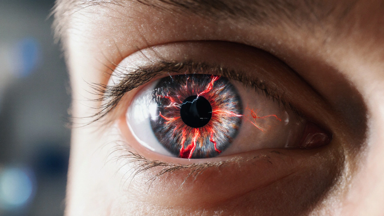
- Reperfusion injury occurs when blood flow returns to oxygen‑starved eye tissue, releasing a burst of harmful free radicals.
- The retina and optic nerve are the most vulnerable structures, and damage can lead to vision loss if untreated.
- Oxidative stress, inflammation, and mitochondrial failure are the three main culprits behind cellular injury.
- Early detection through eye exams and lifestyle tweaks (antioxidant‑rich diet, smoking cessation) can limit permanent damage.
- Research into targeted drugs and gene‑editing approaches is moving fast, offering hope for better neuroprotective therapies.
When blood suddenly rushes back into tissue that has been starved of oxygen, the result can be as nasty as a fireworks display inside cells. This phenomenon, known as reperfusion injury, is especially concerning for the eye because the retina and optic nerve have tiny, high‑demand blood vessels that cannot afford to be flooded with toxic by‑products. Below, we break down what happens, why it matters for your vision, and what you can do right now to protect those precious eyes.
What Is Reperfusion Injury?
Reperfusion injury is a paradoxical tissue damage that follows the restoration of blood flow after a period of ischemia. While re‑oxygenating cells sounds like a cure, the sudden surge of oxygen creates reactive molecules that overwhelm antioxidant defenses, leading to cell death. The term first appeared in cardiac research in the 1960s, but ophthalmologists have since recognized its impact on retinal and optic‑nerve health.
How the Eye Gets Caught in the Reperfusion Loop
Eyes rely on a delicate balance of blood supply: the central retinal artery feeds the inner retina, while the choroidal circulation supports the outer layers. When an artery narrows-due to a clot, vasospasm, or trauma-ischemia sets in. As soon as the blockage clears (spontaneously or via surgery), reperfusion floods the area with:
- Oxygen that fuels reactive oxygen species (ROS).
- Calcium overload that triggers enzymatic cascades.
- Inflammatory cells that release cytokines and proteases.
These events disrupt the blood‑retinal barrier, allowing fluid and immune cells to seep into retinal layers, swelling the tissue and impairing visual signaling.
Early Signs and Clinical Impact
Patients rarely notice the biochemical chaos at first. Typical warning signs include:
- Blurry or patchy vision that appears after a sudden improvement (e.g., after clot‑busting treatment).
- Transient visual field defects, often described as “shadows” or “curtains.”
- Increased eye redness or mild pain, especially if inflammation spreads.
If unchecked, the damage can progress to permanent loss of photoreceptors, thinning of the retinal nerve fiber layer, and optic‑nerve degeneration-conditions that mimic glaucoma or age‑related macular degeneration.

Cellular Players: Oxidative Stress, Inflammation, and Mitochondrial Failure
Three interlinked pathways dominate the cascade:
Oxidative Stress
Oxidative stress arises when ROS outpace the eye’s antioxidant capacity. In the retina, high concentrations of polyunsaturated fatty acids make cell membranes especially vulnerable. When ROS attack, they oxidize lipids, proteins, and DNA, creating a ripple of dysfunction that can trigger apoptosis (programmed cell death).
Inflammation
Within minutes of reperfusion, microglia and infiltrating macrophages release tumor necrosis factor‑α (TNF‑α), interleukin‑1β, and other cytokines. This inflammatory milieu not only harms neurons but also destabilizes the blood‑retinal barrier, making the eye more permeable to harmful substances.
Mitochondrial Dysfunction
Mitochondrial dysfunction is a downstream effect of calcium overload and ROS. Impaired mitochondria produce less ATP, weakening photoreceptor signaling, and release cytochrome c, which further drives cell death pathways.
Protective Strategies: Antioxidants, Pharmacologic Agents, and Lifestyle Tweaks
Because the cascade is rapid, clinicians aim to intervene at multiple points.
Antioxidant Therapy
Compounds such as vitamin C, vitamin E, lutein, zeaxanthin, and the newer molecule N‑acetylcysteine (NAC) have shown promise in animal models. NAC, for instance, replenishes intracellular glutathione, a key detoxifier of ROS, and has moved into early‑phase human trials for retinal ischemia.
Pharmacologic Neuroprotection
Drugs that block calcium channels (e.g., nimodipine) or inhibit inflammatory signaling (e.g., corticosteroids, anti‑TNF agents) can reduce secondary injury. Recent studies on the peptide pigment epithelium‑derived factor (PEDF) suggest it may directly safeguard photoreceptors from oxidative bursts.
Lifestyle Adjustments
Practical steps you can start today:
- Eat a rainbow of fruits and vegetables-spinach, kale, berries, and oranges supply lutein, zeaxanthin, and vitamin C.
- Avoid smoking; tobacco accelerates ROS formation and impairs microvascular health.
- Control blood pressure and diabetes; both conditions raise the risk of ocular ischemia.
- Get regular eye exams that include optical coherence tomography (OCT) to catch early retinal swelling.
Current Research and Emerging Therapies
Scientists are racing to translate bench findings into bedside treatments.
| Mechanism | Retinal Layer | Optic Nerve Impact |
|---|---|---|
| ROS‑mediated lipid peroxidation | Outer nuclear layer (photoreceptors) | Axonal degeneration |
| Inflammatory cytokine surge | Inner plexiform layer | Glial activation |
| Mitochondrial ATP loss | Ganglion cell layer | Reduced nerve conduction |
Clinical trials underway in 2024‑2025 are testing intravitreal injections of PEDF analogs, systemic NAC regimens, and gene‑editing tools that boost endogenous antioxidant enzymes (like superoxide dismutase). Early results show slower retinal thinning and better visual acuity scores compared with standard care.
Practical Tips for Patients and Caregivers
If you or a loved one has experienced an eye‑related ischemic event (e.g., central retinal artery occlusion, ocular stroke, or even severe migraine with visual aura), keep these actions in mind:
- Seek immediate ophthalmic evaluation. Time is tissue; the sooner reperfusion is managed, the better the outlook.
- Ask your eye doctor about antioxidant supplementation, especially if you have risk factors like diabetes.
- Monitor visual changes daily. Use a simple Amsler grid at home to detect subtle distortions.
- Maintain a heart‑healthy lifestyle-regular exercise, low‑sodium diet, and stress reduction-all protect ocular blood flow.
- Stay informed about clinical trials. Registries like ClinicalTrials.gov list recruiting studies on retinal neuroprotection.

Frequently Asked Questions
What triggers reperfusion injury in the eye?
Any sudden restoration of blood flow after a period of blockage-whether from clot‑dissolving medication, surgery, or natural dislodgement-can cause a flood of oxygen‑derived free radicals that damage retinal cells.
Can diet really help prevent damage?
Yes. Foods rich in lutein, zeaxanthin, vitamin C, and omega‑3 fatty acids strengthen the eye’s natural antioxidant defenses, lowering the risk of ROS‑induced injury.
Is there a cure for reperfusion‑related vision loss?
No single cure exists yet, but early intervention with anti‑oxidants, anti‑inflammatory drugs, and emerging neuroprotective agents can halt or even reverse some damage.
How is reperfusion injury diagnosed?
Ophthalmologists use optical coherence tomography (OCT) to spot retinal swelling, fluorescein angiography to assess blood‑flow dynamics, and visual‑field testing to map functional loss.
Are there any promising drugs on the horizon?
Trials of N‑acetylcysteine, PEDF analogs, and gene‑edited antioxidant enzymes are showing encouraging safety and efficacy signals in 2025 studies.


Naresh Sehgal
September 28, 2025 AT 11:14Reperfusion injury is a serious issue, but you can fight it with a proactive mindset. Load up on antioxidant‑rich foods like berries, leafy greens, and nuts – they give your retina the ammo it needs. Stay on top of your blood pressure and blood sugar; the better the circulation, the less of a shock when flow returns. Don’t wait for symptoms – regular OCT scans catch early swelling before it becomes permanent. Keep moving, keep eating clean, and keep your eyes protected.
Kristen Ariies
October 1, 2025 AT 09:44Wow, the cascade of free radicals, calcium overload, and inflammation is like a fireworks show gone wrong, isn’t it?; the retina, with its high‑fuel demand, becomes an easy target for oxidative damage; and the optic nerve, silently watching, can suffer in silence. It’s crucial to pack your diet with lutein, zeaxanthin, and vitamin C, because those antioxidants act like tiny fire extinguishers; they mop up reactive oxygen species before they wreak havoc. Regular eye exams, especially OCT imaging, give you a front‑row seat to any swelling; catching it early can save you from permanent vision loss. And don’t forget lifestyle tweaks-quit smoking, manage blood pressure, and stay active-because a healthy circulatory system is the best defense against reperfusion injury.
Ira Bliss
October 4, 2025 AT 08:14Hey folks 😊! Reperfusion injury can feel like a sudden lightning strike to your retina, but the good news is that a diet rich in leafy greens 🥬, berries 🍓, and omega‑3s can boost your eye’s natural defenses. Adding a daily vitamin C supplement can give your antioxidant levels an extra push. Stay on top of your check‑ups, especially OCT scans, to spot any swelling early. Your eyes will thank you! 🙌
Donny Bryant
October 7, 2025 AT 06:44Reperfusion injury hits the retina hard, so eat foods with lots of vitamins C and E. Keep blood pressure low, and get regular eye check‑ups. Those steps can cut down damage fast.
kuldeep jangra
October 10, 2025 AT 05:14The concept of reperfusion injury may sound like a distant medical term, but its impact on everyday vision is profound, especially for those of us who rely heavily on our sight for work and family. When blood rushes back into an oxygen‑starved retina, the sudden burst of reactive oxygen species behaves like a chemical wildfire, scorching photoreceptor membranes and overwhelming the eye’s natural antioxidant shield. This oxidative onslaught is compounded by a calcium surge that triggers destructive enzymatic pathways, further destabilizing cellular structures. At the same time, inflammatory cells swarm the area, releasing cytokines such as TNF‑α and interleukin‑1β, which not only damage neurons but also compromise the blood‑retinal barrier, allowing fluid to seep in and cause swelling. Mitochondrial dysfunction follows, reducing ATP production and impairing the energy‑hungry retinal ganglion cells, which can lead to irreversible vision loss if not addressed quickly. One of the most effective frontline defenses is a diet saturated with lutein, zeaxanthin, and omega‑3 fatty acids; these nutrients integrate into retinal cell membranes, enhancing resistance to lipid peroxidation. Clinical studies have shown that supplementing with N‑acetylcysteine (NAC) boosts intracellular glutathione levels, giving the eye a stronger line of defense against reactive oxygen species. Regular optical coherence tomography (OCT) appointments enable clinicians to detect subtle retinal thickening before symptoms become noticeable, making early intervention possible. Pharmacologic agents such as calcium channel blockers and anti‑TNF drugs can interrupt the damaging cascades, providing neuroprotection during the critical reperfusion window. Emerging gene‑editing therapies aim to up‑regulate endogenous antioxidant enzymes like superoxide dismutase, promising a future where the eye can self‑repair after ischemic events. Lifestyle factors play a huge role, too; quitting smoking, controlling hypertension, and managing diabetes reduce the baseline risk of vascular occlusions that precipitate reperfusion injury. Exercise improves overall circulatory health, ensuring that blood flow to the retina remains stable and less prone to abrupt changes. It is also vital to avoid excessive exposure to bright lights immediately after an ischemic episode, as this can amplify oxidative stress. For patients who have experienced a central retinal artery occlusion, prompt referral to a retinal specialist can make the difference between partial recovery and permanent blindness. In summary, a multi‑pronged approach-nutritional, pharmacologic, lifestyle, and vigilant monitoring-offers the best chance to mitigate the devastating effects of reperfusion injury on the eye.
harry wheeler
October 13, 2025 AT 03:44Antioxidants are your best bet against reperfusion damage.
faith long
October 16, 2025 AT 02:14The cascade you described is a perfect storm for the eye, and I’ve seen patients who didn’t act fast end up with permanent vision loss. Getting on a high‑dose antioxidant regimen early, especially NAC, can actually neutralize a lot of those free radicals before they do serious harm. Pair that with a short course of anti‑inflammatory steroids, and you’re hitting both the oxidative and inflammatory arms of the injury. Don’t forget to monitor intra‑ocular pressure; inflammation can spike it, risking further damage. And always push for OCT imaging – it’s the clearest window into what’s happening at the cellular level.
Danny Wakefield
October 19, 2025 AT 00:44Sounds like the eye’s version of a bomb blast; good thing we have some armor now.
Randy Faulk
October 21, 2025 AT 23:14From a pathophysiological standpoint, reperfusion injury exemplifies the paradox wherein restoration of oxygenated blood precipitates a secondary wave of cellular demise. The intricate interplay among reactive oxygen species, calcium overload, and pro‑inflammatory cytokines constitutes a triad that overwhelms retinal resilience. Empirical evidence advocates for the prophylactic administration of antioxidants such as N‑acetylcysteine, which replenishes glutathione reservoirs, thereby attenuating oxidative insult. Moreover, targeted anti‑inflammatory agents, including corticosteroids and novel anti‑TNF biologics, have demonstrated efficacy in preserving retinal architecture. A vigilant surveillance regimen employing optical coherence tomography remains indispensable for early detection and therapeutic titration.
Brandi Hagen
October 24, 2025 AT 21:44💥 Wow, the eye’s response to sudden blood flow is like a battlefield, and we can’t let those free radicals win! 🚀 Load up on lutein, zeaxanthin, and vitamin C – they’re the frontline troops protecting our vision. 🛡️ Don’t wait for symptoms; demand OCT scans and aggressive antioxidant therapy now. 👊 Our eyes deserve the best defense, and we have the science to back it up! 🌟
isabel zurutuza
October 27, 2025 AT 19:14Oh great, another reminder to eat kale and get an OCT.
James Madrid
October 30, 2025 AT 17:44Great rundown! Adding a daily dose of omega‑3 fish oil can further bolster retinal health, and keeping blood sugar in check helps prevent those ischemic spikes. Keep sharing these insights.
Justin Valois
November 2, 2025 AT 16:14Reperfusion injury is like a firecracker in your retina – BOOM! If you dont stack up on antioxidants, you're basically signing up for fast vision loss. Get those lilac greens, berries, and maybe a NAC supplement. And dont forget the doc when you see any weird shadows.
Jessica Simpson
November 5, 2025 AT 14:44I was curious about how the blood‑retinal barrier gets compromised, and it turns out the inflammation really opens the doors for fluid. It’s fascinating that lifestyle tweaks can actually close those doors again. Definitely worth a deeper dive.
Ryan Smith
November 8, 2025 AT 13:14Sure, just sprinkle some antioxidants and the eye will be fine – as if it’s that simple.
John Carruth
November 11, 2025 AT 11:44This post nails the multi‑layered nature of reperfusion injury, and I think we can all agree that early detection is the linchpin of any effective intervention. By coupling antioxidant therapy with anti‑inflammatory steroids, we’re essentially attacking both the fire and the smoke at once. Adding lifestyle changes, like a Mediterranean diet rich in leafy greens and omega‑3s, creates a sustainable defense that supports long‑term retinal health. Regular OCT monitoring provides the feedback loop necessary to adjust treatment before permanent damage sets in. The emerging gene‑editing approaches sound promising, especially those boosting endogenous superoxide dismutase, but we should stay grounded until larger trials confirm safety. Overall, a proactive, layered strategy gives patients the best shot at preserving vision.
Melodi Young
November 14, 2025 AT 10:14Nice summary, very helpful!